Human anatomy and physiology lab manuals are crucial for a hands-on learning experience‚ often featuring versions for Pearson‚ pig‚ and cat dissections.
These manuals‚ like the 10th edition by Pearson‚ provide detailed exercises and support materials for effective lab quizzing and understanding anatomical structures.
They guide students through dissections‚ microscopic observations‚ and physiological measurements‚ enhancing comprehension of the human body’s complex systems.
Purpose of Laboratory Manuals
Human anatomy and physiology laboratory manuals serve as essential companions to classroom learning‚ bridging theoretical knowledge with practical application. Their primary purpose is to provide structured‚ hands-on experiences that reinforce concepts and develop critical thinking skills.
These manuals‚ such as those offered by Pearson and versions utilizing pig or cat specimens‚ guide students through dissections‚ microscopic examinations‚ and physiological experiments. They facilitate a deeper understanding of anatomical structures‚ functional relationships‚ and experimental design.
Furthermore‚ they often include pre-lab exercises‚ observation prompts‚ and post-lab questions to encourage active learning and assessment. The 10th edition and other resources aim to enhance lab quizzing and overall comprehension of the human body’s intricate systems‚ fostering a robust foundation in the biological sciences.
Importance of Hands-on Learning
Hands-on learning is paramount in human anatomy and physiology‚ as it transcends rote memorization and fosters a deeper‚ more lasting understanding. Laboratory manuals‚ including Pearson’s latest editions and pig/cat versions‚ facilitate this crucial experiential learning.
Dissection‚ microscopic observation‚ and physiological measurements allow students to directly interact with anatomical structures and processes‚ solidifying theoretical concepts. This active engagement enhances retention and develops critical thinking skills essential for future scientific endeavors.
The ability to apply knowledge in a practical setting‚ guided by detailed instructions and assessments within the lab manual‚ is invaluable. Mastering A&P course materials and dynamic study modules further support this‚ ensuring a comprehensive and effective learning experience.
Essential Laboratory Equipment
Essential tools include microscopes for cellular study‚ dissection kits for anatomical exploration‚ and safety gear‚ all detailed within comprehensive human anatomy lab manuals.
The Microscope: Principles and Use
Microscopes are foundational tools in anatomy and physiology labs‚ enabling visualization of cells and tissues beyond the naked eye’s capability. Lab manuals‚ such as those from Pearson‚ dedicate significant sections to microscopic principles.
Understanding magnification‚ resolution‚ and proper illumination techniques is crucial. Students learn to prepare slides‚ focus specimens using objective lenses‚ and identify cellular structures.
The manuals detail various microscope types – brightfield‚ phase contrast‚ and potentially electron microscopes – explaining their specific applications. Proper handling and maintenance are emphasized to ensure longevity and accurate observations.
Exercises often involve observing prepared slides of different tissue types‚ reinforcing histological concepts and developing observational skills.
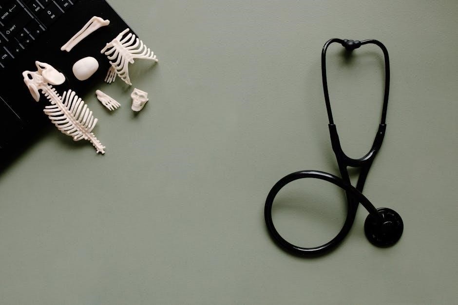
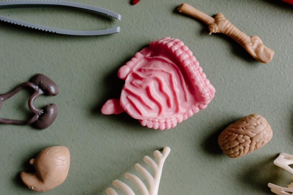
Dissection Tools and Techniques
Dissection is a cornerstone of anatomy labs‚ requiring specialized tools and precise techniques. Laboratory manuals‚ including pig and cat versions‚ provide detailed guidance on safe and effective dissection procedures.
Essential tools include scalpels‚ scissors‚ probes‚ forceps‚ and pins‚ each serving a specific purpose in revealing anatomical structures. Manuals emphasize proper handling and safety protocols to prevent injury.
Students learn systematic dissection approaches‚ carefully separating layers of tissue to expose muscles‚ nerves‚ and organs. Accurate identification and labeling are crucial skills developed through these exercises.
The manuals often include diagrams and illustrations to aid in visualization and understanding of anatomical relationships‚ enhancing the learning experience.
Safety Protocols in the Anatomy Lab
Safety is paramount in the anatomy lab‚ and laboratory manuals dedicate significant attention to establishing and enforcing strict protocols. These guidelines protect students and ensure a respectful learning environment.
Essential safety measures include wearing appropriate personal protective equipment (PPE) – gloves‚ goggles‚ and lab coats – to prevent exposure to specimens and chemicals.
Proper handling and disposal of dissection tools‚ especially scalpels‚ are emphasized to avoid accidental injuries. Manuals detail procedures for handling biological specimens‚ including proper preservation and disposal methods.
Students are instructed on chemical safety‚ including the safe use and disposal of preservatives and cleaning agents. Adherence to these protocols is crucial for a safe and productive lab experience.
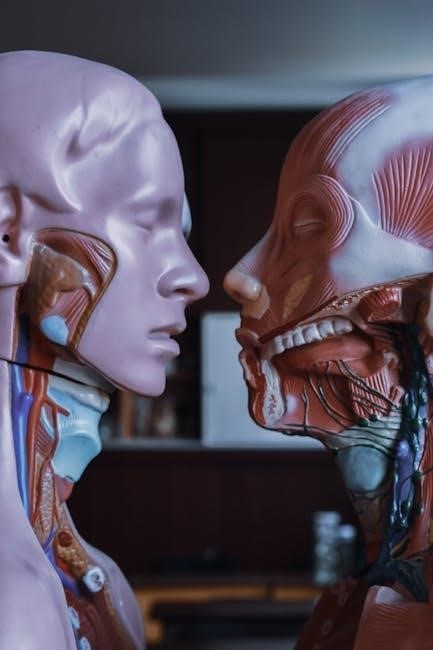
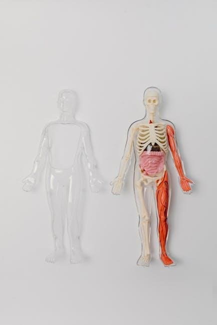
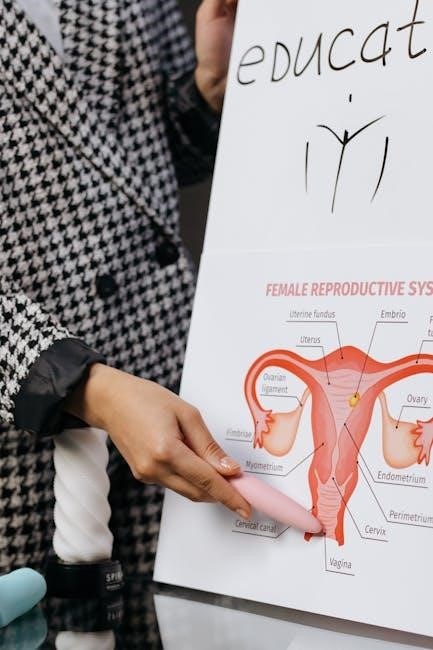
Body Organization and Anatomical Terminology
Lab manuals introduce the language of anatomy‚ including anatomical position‚ directional terms‚ body planes‚ and cavities‚ for precise communication.
Anatomical Position and Directional Terms
Human anatomy lab manuals emphasize the standardized anatomical position – standing erect‚ feet slightly apart‚ palms facing forward – as a foundational reference point.
Understanding directional terms is paramount; manuals meticulously define superior (above)‚ inferior (below)‚ anterior (front)‚ posterior (back)‚ medial (midline)‚ and lateral (side) relationships.
These terms enable precise description of structures and their locations relative to one another‚ crucial for dissection and accurate communication.
Lab exercises often require students to identify these directional relationships on anatomical models or cadaver specimens‚ solidifying their comprehension.
Mastering this terminology is essential for navigating the complexities of human anatomy and physiology effectively.
Body Planes and Cavities
Human anatomy lab manuals detail the three primary body planes: sagittal (midline‚ dividing left/right)‚ frontal (dividing anterior/posterior)‚ and transverse (dividing superior/inferior).
Understanding these planes is vital for interpreting anatomical sections and imaging techniques‚ like those encountered in lab exercises.
Manuals also thoroughly explain the major body cavities – dorsal (cranial & vertebral) and ventral (thoracic‚ abdominal‚ & pelvic) – and their contents.
Students learn to correlate specific organs with their respective cavities‚ enhancing spatial understanding of anatomical organization.
Lab activities often involve identifying structures within these cavities on models or diagrams‚ reinforcing the relationship between plane‚ cavity‚ and organ location.
Histology: The Study of Tissues
Human anatomy & physiology lab manuals dedicate significant sections to histology – the microscopic study of tissues.
Students learn to identify the four primary tissue types: epithelial‚ connective‚ muscle‚ and nervous‚ recognizing their unique structures and functions.
Lab exercises commonly involve examining prepared slides under a microscope‚ differentiating tissues based on cellular arrangement and matrix composition.
Manuals provide detailed illustrations and descriptions aiding in accurate tissue identification‚ crucial for understanding organ structure and function.
Understanding histological features is fundamental‚ as tissues form the building blocks of all organs and systems explored throughout the lab course.

System-Specific Laboratory Exercises
Lab manuals guide dissections and identifications within each body system – skeletal‚ muscular‚ and nervous – enhancing anatomical understanding through practical application.

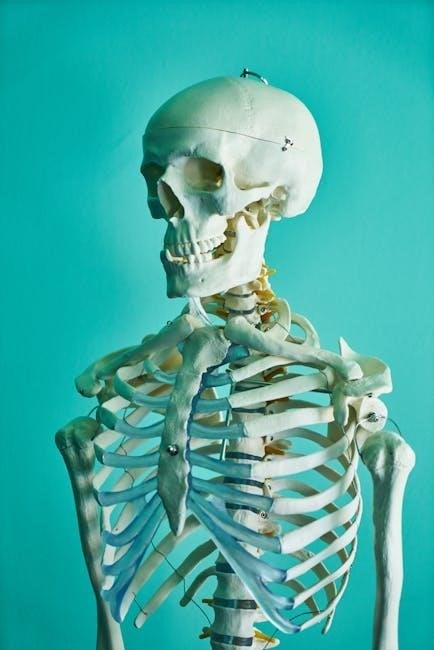
Skeletal System: Bone Identification and Structure
Laboratory exercises focusing on the skeletal system are fundamental to understanding human anatomy. These typically involve meticulous bone identification‚ requiring students to differentiate between various bones – cranial‚ vertebral‚ appendicular – based on their unique morphological features.
Students learn to identify key bone structures like processes‚ foramina‚ and fossae‚ understanding their functional significance. Manuals often include diagrams and illustrations to aid in accurate identification.
Practical application extends to examining bone tissue under a microscope‚ observing compact and spongy bone arrangements. Furthermore‚ exercises may involve articulating bones to demonstrate joint types and range of motion‚ solidifying comprehension of skeletal system mechanics and overall body support.
Muscular System: Muscle Identification and Function
Laboratory exercises dedicated to the muscular system emphasize both accurate muscle identification and a thorough understanding of their respective functions. Students utilize laboratory manuals to locate superficial muscles‚ learning origins‚ insertions‚ and actions through palpation and dissection – often utilizing pig or cat models.
Exercises frequently involve identifying muscle fiber arrangements and relating them to contractile strength and range of motion. Manuals often incorporate diagrams illustrating muscle attachments and lever systems.
Furthermore‚ labs may include electromyography (EMG) to demonstrate muscle activity during contraction‚ linking anatomical knowledge with physiological processes. This hands-on approach reinforces comprehension of how muscles contribute to movement and overall body function.
Nervous System: Brain Dissection and Nerve Identification
Laboratory manuals guide students through detailed brain dissection‚ enabling identification of major brain regions – cerebrum‚ cerebellum‚ and brainstem – and their associated functions. Pig or cat brains are commonly used for this purpose‚ providing a tangible learning experience.
Exercises focus on recognizing cranial nerves‚ tracing their pathways‚ and correlating them with sensory and motor functions. Students learn to differentiate between gray and white matter‚ and identify key structures like the spinal cord.
Manuals often include diagrams and illustrations to aid in nerve identification and understanding neural pathways. This hands-on exploration solidifies comprehension of the nervous system’s complex organization and its role in coordinating bodily functions.
Physiological Measurements and Analysis
Lab manuals detail techniques for measuring heart rate‚ blood pressure‚ lung capacity‚ and utilizing electromyography (EMG) to analyze muscle activity.
Heart Rate and Blood Pressure Measurement
Laboratory manuals dedicate significant sections to understanding cardiovascular physiology‚ emphasizing practical heart rate and blood pressure measurement techniques. Students learn to accurately assess pulse rates at various locations‚ correlating them with physiological states like exercise.
Detailed protocols guide the use of a sphygmomanometer to determine systolic and diastolic blood pressure‚ crucial indicators of cardiovascular health.
Exercises often involve analyzing the effects of posture‚ activity‚ and even psychological stress on these vital signs.
Furthermore‚ manuals explain the underlying mechanisms regulating blood pressure‚ linking anatomical structures – like arteries and the heart – to their functional roles.
Data analysis and interpretation are key components‚ fostering a deeper understanding of cardiovascular dynamics.
Lung Capacity and Respiratory Rate
Human anatomy & physiology laboratory manuals provide comprehensive exercises focused on respiratory system function‚ specifically lung capacity and respiratory rate. Students utilize spirometers to measure vital capacities‚ including tidal volume‚ inspiratory reserve volume‚ and expiratory reserve volume.
These measurements are then used to calculate total lung capacity‚ offering insights into individual respiratory health.
Protocols detail methods for accurately determining resting respiratory rate and observing changes during exercise.
Manuals explain the anatomical basis of these measurements‚ connecting lung volumes to structures like alveoli and the diaphragm.
Data analysis emphasizes interpreting results in relation to factors like age‚ sex‚ and physical activity level.
Electromyography (EMG) and Muscle Activity
Human anatomy & physiology laboratory manuals incorporate electromyography (EMG) to investigate muscle activity. These exercises demonstrate how EMG records the electrical activity produced by skeletal muscles during contraction.
Students learn to properly place electrodes on specific muscles and correlate EMG signal amplitude with varying levels of muscle force.
Manuals detail protocols for assessing muscle fatigue and the effects of different stimuli on muscle recruitment.
The connection between nerve impulses and muscle fiber activation is emphasized‚ linking physiological principles to observable EMG patterns.
Data analysis focuses on interpreting EMG waveforms and understanding their relationship to muscle function and neurological control.
Laboratory Manual Versions & Resources
Human anatomy & physiology labs offer diverse resources‚ including Pearson editions‚ pig versions‚ and cat dissections‚ catering to varied instructional needs.

Pearson Human Anatomy & Physiology Lab Manual
Pearson’s Human Anatomy & Physiology Lab Manual stands as a leading resource‚ frequently updated with new editions like the 10th‚ identified by ISBNs 9781292026374 and 9781292038766.
This widely adopted manual excels due to its extensive instructor support materials‚ designed to simplify the creation of lab quizzes and assessments.
The table of contents systematically covers foundational concepts‚ beginning with “The Language of Anatomy” and progressing to detailed explorations of microscopic techniques using “The Microscope.”
It provides a structured approach to learning‚ guiding students through practical exercises that reinforce theoretical knowledge.
The manual’s comprehensive nature makes it a valuable tool for both students and educators in anatomy and physiology courses.
Pig Version Laboratory Manuals
Pig version laboratory manuals‚ such as Melissa Greene’s “Laboratory Manual for Human Anatomy & Physiology: A Hands-on Approach‚” offer a cost-effective and ethically considerate alternative to human cadaver dissection.
These manuals utilize the pig as a model organism due to its significant anatomical similarities to humans‚ allowing students to explore organ systems and structures in a tangible way.
Available in downloadable PDF format‚ these resources provide detailed instructions for dissection‚ identification of anatomical features‚ and understanding physiological functions.
The hands-on approach fosters a deeper comprehension of anatomy and physiology‚ enhancing learning beyond traditional textbook study.
These manuals are designed to complement coursework and provide a practical‚ engaging laboratory experience for students.
Cat Version Laboratory Manuals
Cat version laboratory manuals‚ like the 12th Edition by Marieb and Smith‚ remain a classic choice for anatomy and physiology education‚ providing a detailed exploration of mammalian anatomy.
These manuals facilitate in-depth dissection exercises‚ enabling students to identify and understand the relationships between various organ systems within a complex organism.
The ISBN 0321971353 and 978-0321971357 identify this widely used resource‚ often available in PDF format for convenient access and study.
Students formulate and test hypotheses during experiments‚ strengthening their scientific reasoning skills alongside anatomical knowledge.
These manuals offer a comprehensive learning experience‚ bridging theoretical concepts with practical application through detailed anatomical investigation.